Geoff Orbell
Clinical history
Lucy, a seven-year-old, spayed female Domestic Short-Haired cat presented to her veterinarian with a palpable subcutaneous mass in the jugular furrow on the left side of her neck. The owners opted for surgical excision and during surgery it was noted to be adherent to the adventitia of the underlying jugular vein. The mass was successful removed and the cat recovered uneventfully.
Laboratory findings
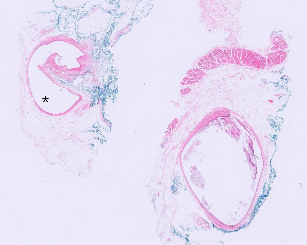
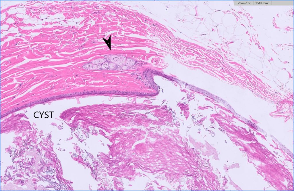
Histologically there were two adjacent cysts which were lined by different epithelia (Figure 1). One cyst was lined by stratified squamous epithelium and contained abundant laminated keratin (Figure 2). Rare peripheral sebaceous glands in the surrounding stroma were identified communicating with the cyst which confirmed this was a dermoid cyst.


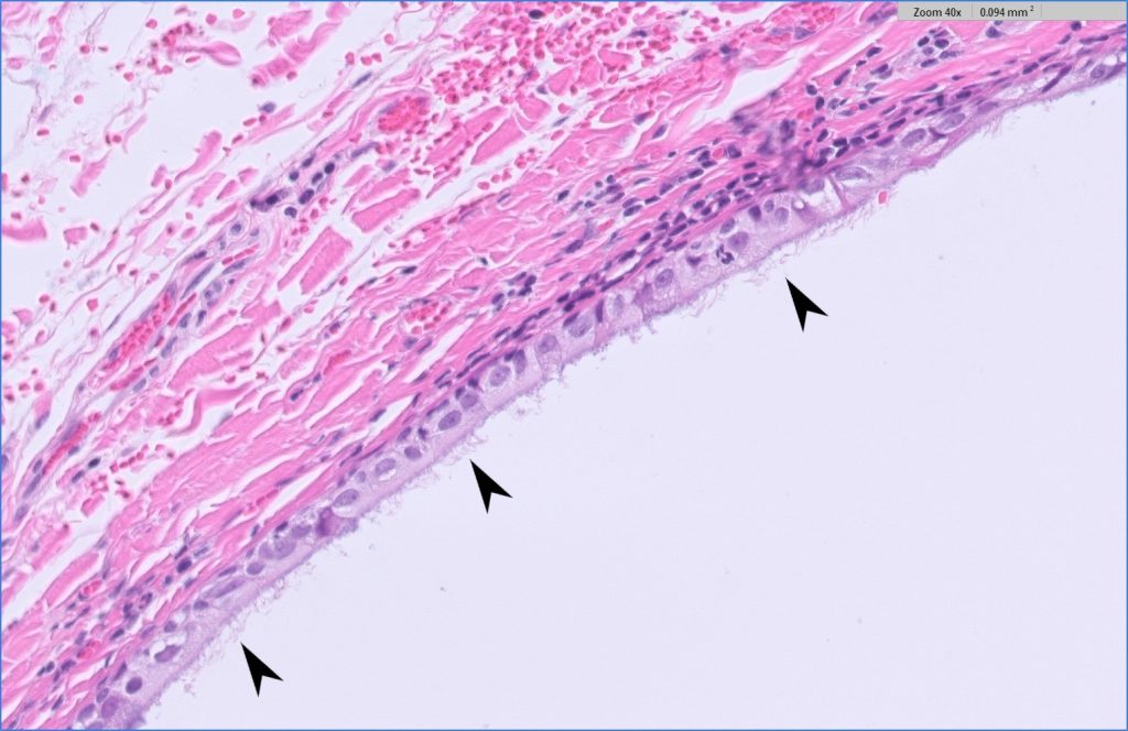
The second cyst (Figure 3) contained clear fluid and was lined by a solitary layer of columnar epithelium which was ciliated consistent with a branchial pouch cyst.

Discussion
True cysts are defined as epithelial-lined structures with no external opening. The most common one we see in veterinary medicine are epidermal inclusion or follicular cysts which are usually acquired due to some obstruction of the hair follicle which continues to produce keratin.
Dermoid cysts are similar to follicular cysts histologically but are usually identified in young animals less than one-year old, as they form early in gestation. They develop due to sequestration of skin ectoderm during embryogenesis, most commonly due to failure of ectoderm to fully separate from the neural tube, which is why some dermoid cysts can communicate with the spinal cord forming dermoid sinuses (e.g. Rhodesian Ridgebacks).
Dermoid cysts are much more common in dogs than cats and occur most commonly along the dorsal midline (including tail) and are often multiple and linearly arranged. Dermoid cysts in cats are much rarer and have been reported in the subcutis and retropharynx.
Branchial pouch cysts are much rarer in veterinary species but are also benign congenital lesions derived from different embryonic structures (branchial arches) than dermoid cysts. Histologically they can appear similar to dermoid cysts with keratinizing squamous epithelium but they lack associated adnexa. A proportion of them are lined with ciliated columnar epithelium which is a pathognomonic feature given their location.
Other congenital cysts derived from the branchial arches seen in veterinary medicine include thyroglossal cysts, ultimobranchial cysts and thymic cysts.
Dermoid and branchial pouch cysts are benign, slow growing lesions and will not recur if completely excised.
Acknowledgements to Ana at Kelburn Vets and Lucy’s owner who gave us permission to publish this case.
Cover photo credit: sabri-tuzcu-unsplash

