Alice Fraser
Bovine adenovirus raises its ugly head each autumn and winter in young cattle, 6 to 12 months of age (predominantly rising one year olds/R1s). Clinical presentation is usually acute scours (watery to haemorrhagic) and rapid death, or sudden death without preceding signs noticed. Occasionally, respiratory signs are noted. Usually a few animals in a mob are affected.
Awanui Veterinary laboratories saw several cases of this disease during the recent season, both in the North and South Islands.
Case 1
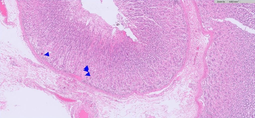
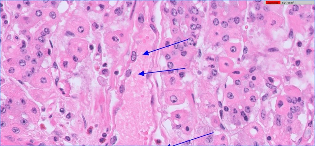
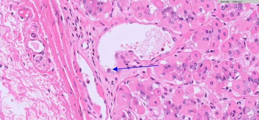
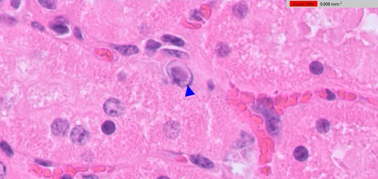
A group of thirty R1s, three animals died following an acute bout of severe scours. Post mortem tissues from one animal were submitted to the laboratory, including abomasum, small intestine, liver and kidney. Histologic evaluation of the abomasum revealed a haemorrhagic necrotising abomasitis. In addition, multifocal mucosal blood vessels revealed endothelial basophilic intranuclear inclusions (Figures 1, 2 and 3). Significant submucosal oedema results from the vascular pathology. These changes, notably with the evidence of intranuclear inclusion bodies, are characteristic of the disease.
Case 2
Following a spell of inclement weather, two out of a group of R1 dairy heifer grazers, which had previously shown their expected liveweight gain, were found recumbent and moribund with evidence of acute scours. Post mortem tissues were harvested and formalin-fixed, including abomasum, intestines, kidney and liver. Histologic evaluation revealed large basophilic intranuclear adenoviral inclusions within the vascular endothelial cells in the kidney, abomasum, small and large intestines. Figure 4 shows these prominent inclusion bodies present in the renal vascular endothelial cells. In addition, there was a mild multifocal lymphoplasmacytic/neutrophilic interstitial nephritis. Other changes included acute haemorrhagic abomasitis (similar to the above case) and acute haemorrhagic enterocolitis. In this case, a PCR test for Bovine Adenovirus on faeces was carried out simultaneously giving a positive result.
Outbreaks
Bovine adenovirus infections typically cause small outbreaks of acute enteric disease and deaths in young cattle during autumn and winter (sometimes into spring depending on the season). Occasionally respiratory signs are a feature. Outbreaks are usually preceded by a stress factor or factors, including inclement weather, feed shortages, recent transport, concomitant parasitic burdens or infections.
Previous serologic surveys show widespread seroconversion to adenovirus in cattle populations throughout New Zealand; it in as yet unclear why some animals develop severe systemic infections. Several different adenovirus types have been detected in cattle, although BAdV-10 is implicated in nearly all clinical cases where qPCR confirmation is undertaken (Vaatstra et al, 2016). The PCR test is predominantly carried out on a faecal sample. Differential diagnoses for acute severe enteric disease/sudden death in this age group of calves includes Salmonellosis, Yersiniosis, mucosal disease, gastroenteric parasites, toxicosis (e.g. plant nitrates, arsenic), clostridial deaths.
Reference:
Vaatstra BL, Tisdall DJ, Blackwood M and Fairley RA. Clinicopathological features of 11 suspected outbreaks of Bovine Adenovirus infection and development of a real-time quantitative PCR to detect Bovine Adenovirus type 10. NZVJ. 64:308-313, 2016.

