
Kathryn Jenkins
Reticulocyte haemoglobin (RET-He) is a valuable diagnostic marker for iron-deficient erythropoiesis in both dogs and cats. RET-He reflects current iron availability and enables earlier identification of iron-limited states.
Reticulocytes are immature red blood cells (erythrocytes), which develop in the bone marrow and are released into circulation prior to maturation. Maturation time in dogs takes around two days, whilst in cats aggregate reticulocytes mature within one day (leaving maturing punctate reticulocytes to circulate for a further 2 weeks). In routinely stained blood films, most reticulocytes appear as polychromatophils due to the RNA staining a diffuse blue. Special stains (e.g. New Methylene Blue, or analyser fluorescent stains) are used to accurately count reticulocytes, see Figure 1.
Iron deficiency can be either absolute or functional. Absolute iron deficiency occurs with decreased iron stores such as chronic blood loss (most common cause in dogs), or nutritional deficiency (uncommon in veterinary medicine). Functional iron deficiency occurs with normal iron body stores, but reduced availability of iron for erythropoiesis. This can occur with either anaemia of inflammation/chronic disease (most common cause in cats), or with portosystemic shunts (due to inadequate protein synthesis required for iron metabolism).
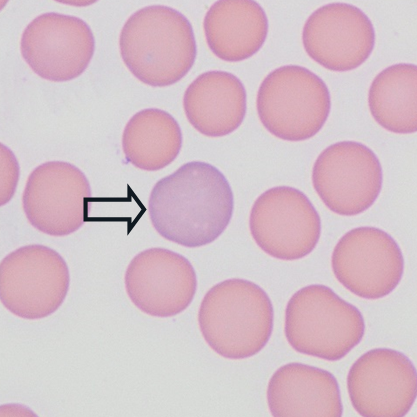
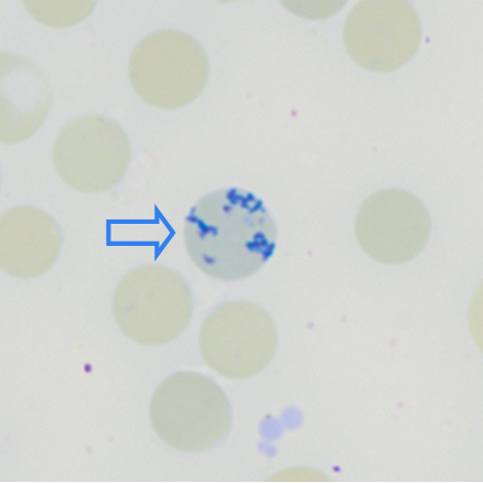
Figure1: Immature erythrocytes. A polychromatophil (black arrow) in a routine stain (appears larger and more basophilic than mature erythrocytes). This cell would be called a reticulocyte if we use either a NMB stain (blue arrow) which causes the RNA to clump in a reticular pattern, or with fluorescent stains that are used in some haematology analysers.
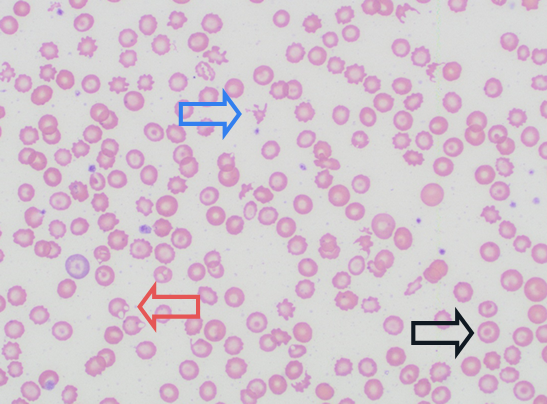
Iron deficiency anaemia secondary to chronic blood loss occurs as a consequence of both haemorrhage and reduced life span of fragile red blood cells. The characteristic morphologic changes of iron deficiency anaemia occur with chronicity, due to the relatively long lifespan of erythrocytes (around 100 days in dogs, and 80 days in cats). Initially, iron deficiency anaemia appears regenerative, and with time presents as small (microcytic) erythrocytes with increased central pallor (hypochromic, target cells) and morphologic changes associated with increased fragility (e.g. schistocytes, keratocytes) see Figure 2. Decreased RBC indices (MCV, MCH and MCHC) are seen in advanced cases, but are often within reference in early stages as they are insensitive to changes in small proportions of erythrocytes.
Anaemia of inflammatory disease occurs as a consequence of suppression of erythropoiesis (via inflammatory cytokines), reduced iron availability (via IL-6 upregulation of hepcidin), and extravascular haemolysis. These cases often present with unremarkable appearing erythrocytes on blood film review (i.e. normochromic, normocytic). However, with protracted cases, anaemia of inflammatory disease may also present with microcytic hypochromic erythrocytes. PCV in these cases is usually not lower than 20%, and poikilocytosis (such as schistocytes, keratocytes) are not typical. There may be an inflammatory leukogram and protein changes may include decreased albumin with increased globulin.
It is also possible to have cases of combined blood loss and inflammation.
RET-He content is decreased with both absolute and functional iron deficiency, and allows earlier detection of clinical cases prior to morphologic changes being detectable on the blood film or notable decreases in MCV, MCH and MCHC.
RET-He is measured by reference laboratory analysers (including the Sysmex XN used at Awanui Veterinary), and the ProCyte Dx in-house analyser. In cases of iron-deficient erythropoiesis, RET-He level is expected to be decreased. As reticulocytes circulate for a short time in peripheral blood, decreased RET-He is an early indicator of iron deficient erythropoiesis.
RET-He may appear falsely increased with macrocytosis, or falsely decreased in rare cases presenting with microcytosis without hypochromasia (including breed related microcytosis in Akita and Shiba Inu, Chow Chow or Shar Pei). RET-He appears stable over 48 hours, however analysis of fresh blood is recommended regardless due to effects of storage artefact on other parameters.
Published studies suggest a RET-He below 20.9 pg supports iron deficient erythropoiesis in dogs, and below 12.5 pg supports iron deficient erythropoiesis in cats. Further research is required to see if RET-He levels can help differentiate absolute vs functional iron deficiency.
In cases of anaemia in clinical practice, a blood film review is an essential component of a full CBC work-up. Your local Awanui Veterinary pathologists are always here to help.
References:
· Keiner et al. Evaluation of reticulocyte hemoglobin content (RETIC-HGB) for the diagnosis of iron-limited erythropoiesis in cats. Veterinary Clinical Pathology. 49, 2020.
· Fuchs et al. Canine reticulocyte hemoglobin content (RET-He) in different types of iron-deficient erythropoiesis. Veterinary Clinical Pathology. 46/3, 2017.
· Fuchs et al. Evaluation of reticulocyte hemoglobin content (RET-He) in the diagnosis of iron-deficient erythropoiesis in dogs. Veterinary Clinical Pathology. 46/4, 2017.