

Lisa Hulme-Moir
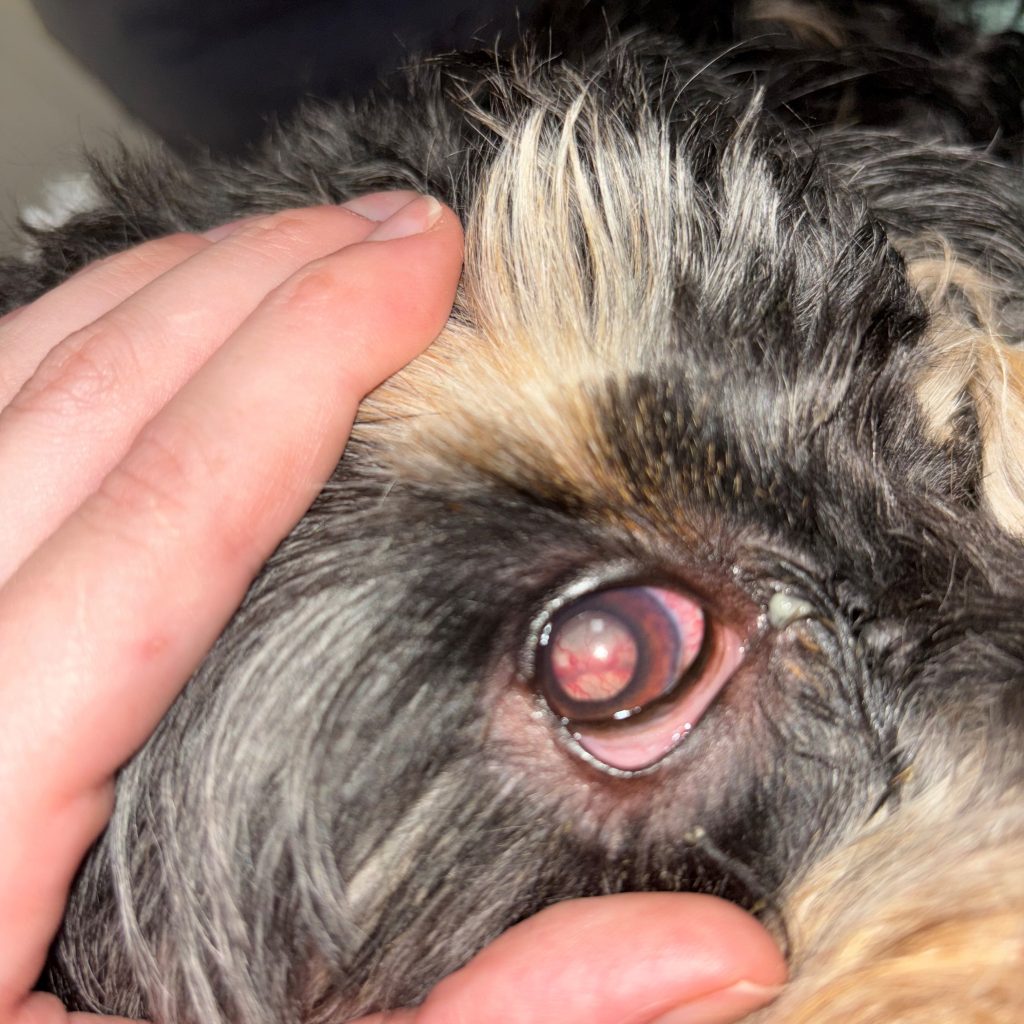
A four-year old male neutered Cavoodle was presented to a referral clinic with a three-month history of haemorrhagic colitis that had not responded to treatment with antiparasitics, antibiotics or dietary modification. He had been started on corticosteroids but over the weekend had developed acute blindness in one eye due to exudative retinal detachment and glaucoma (figure 1) with emerging changes in the other eye. Enucleation of eye was performed and was submitted to the laboratory along with vitreous humour collected pre-fixation, fresh and fixed colonic biopsies and rectal smears for cytology.
Laboratory testing
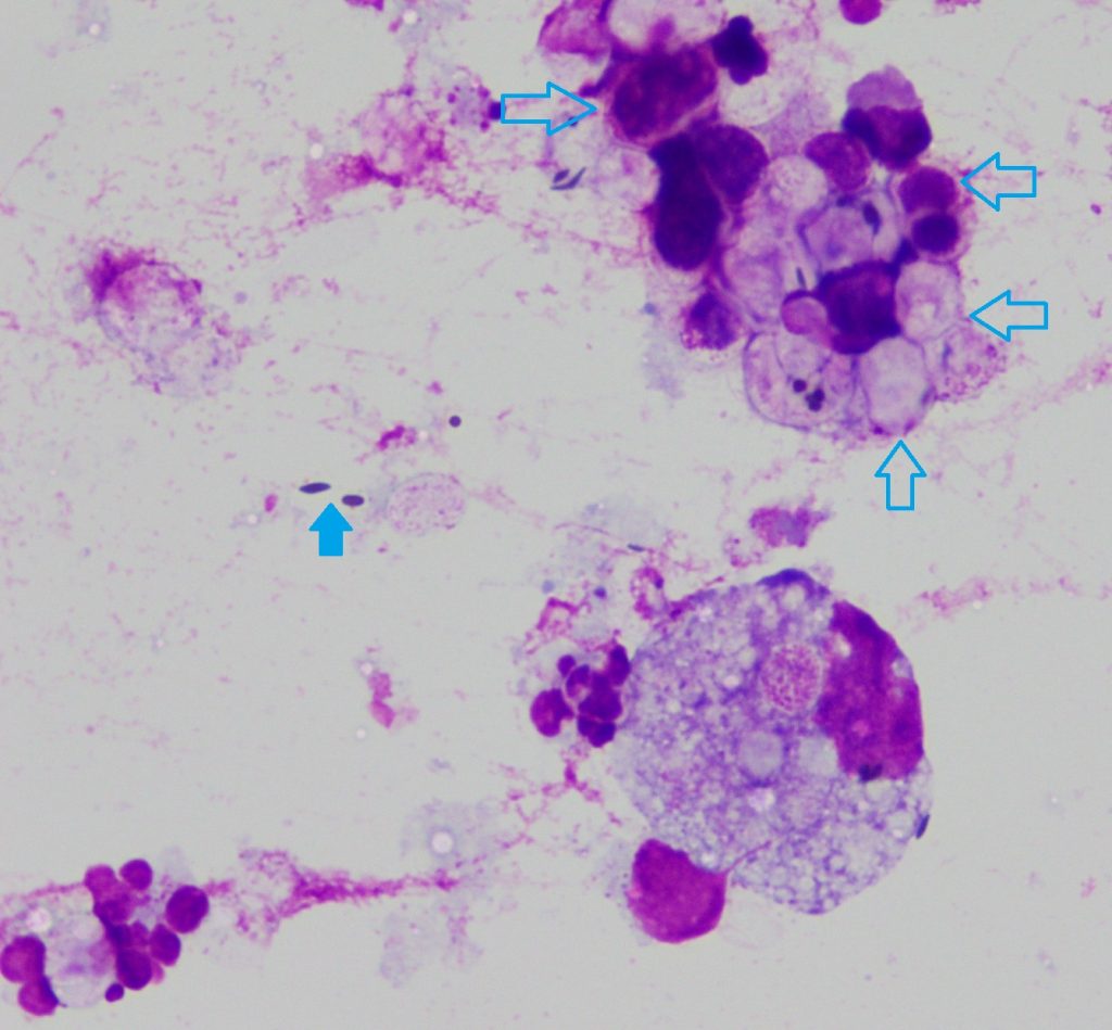
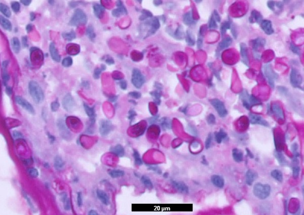
Cytology performed on cytocentrifuged preparations of the vitreous humour revealed mixed inflammation with numerous organisms consistent with Prototheca sp. (figure 2).
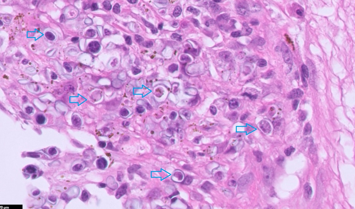
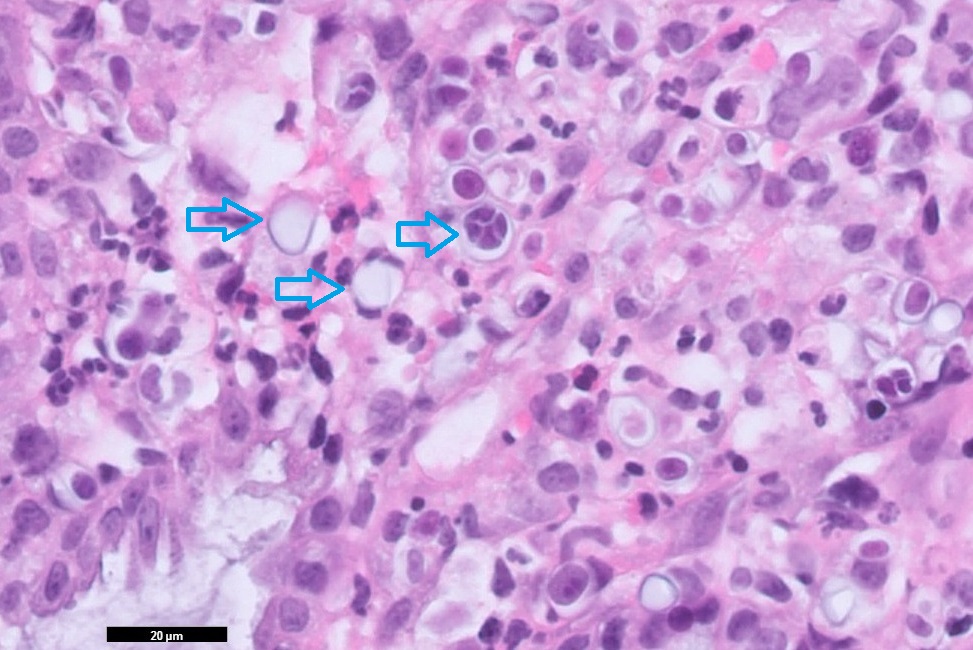
Examination of tissue sections of the enucleated eye and colon also detected numerous algae (figure 3) and culture of the colonic biopsies yielded a heavy growth of Prototheca sp. further confirming the diagnosis of protothecosis. Prototheca were not detected in the rectal smears.
Discussion
Prototheca are achlorophylic algae that are found widely in the environment, particularly damp habitats rich in organic material. As they lack chloroplasts, they are unable to photosynthesize and thus rely on external sources for their nutrients. Although an infrequent cause of disease, protothecosis is most commonly diagnosed in cattle where they can cause sporadic cases and outbreaks of mastitis in herds and occasionally skin infections. They are also capable of causing skin and systemic infections in humans and other animal species.
In dogs, infection typically starts in the large intestine and cases very commonly have a history of chronic large bowel diarrhoea followed by sudden clinical signs attributable to disseminated disease. This most commonly manifests as acute blindness and retinal detachment due to ocular involvement, but neurologic signs and urinary tract signs can also occur and on postmortem, there is typically evidence of infection in multiple organs (Stenner et al. 2007). Cutaneous infection has also been reported in dogs, which may be due to haematogenous spread or local inoculation of the organism.
In New Zealand, there have been several recent case reports of protothecosis in dogs (Walker et al 2022, Price et al 2023) and this disease should be considered as a differential diagnosis for any dogs with a history of chronic diarrhoea presenting with acute ocular or neurologic signs.
References
- Price et al. Protothecosis in four dogs in New Zealand. New Zealand Veterinary Journal. 71:321-328, 2023.
- Stenner et al. Protothecosis in 17 Australian dogs and a review of the canine literature. Medical Mycology. 45:249-266, 2007.
- Walker et al. Disseminated protothecosis with central nervous system involvement in a dog in New Zealand. New Zealand Veterinary Journal. 70:238-243, 2022.