Species: All.
Collection protocol:
Skin disease can be frustrating in clinical practice, but it should be remembered that the skin is one of the few organs that is easily available for examination. By careful systematic observation, the diagnoses of many dermatoses can be determined, or at least a differential list established. Specialist dermatologists will often diagnose and treat animals on the basis of the history, signalment, physical examination and a few ancillary tests (e.g. cytology).
It is important that all of the skin and the external mucous membranes are examined. Recognising the morphology of skin lesions is essential in diagnosing skin problems. Often primary lesions (e.g. vesicles) are obscured by secondary ones (e.g. crusts). Changes due to medication and self-induced trauma are also common secondary problems encountered in examination.
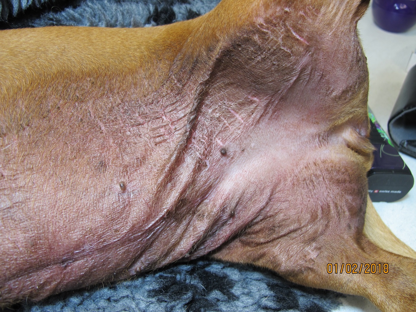
It is helpful for the pathologist if skin lesions and their distribution are accurately described on the accession form. It’s also very helpful to indicate on the drawing from which anatomical area the biopsies were taken. The signalment, previous history and previous treatments of the case are critical in diagnosing skin disease, and we can help you much more armed with that information. Your clinical differential diagnoses are also always important. Do not worry about introducing “bias” into our process; we cannot see lesions that are not there, and rather than influencing us in the wrong direction, it is much more likely that your clinical information will allow us to provide specific and accurate interpretations.
Digital pictures are very useful as another means of submitting extra information to us. Please contact Awanui Veterinary for the email address of a pathologist to send them to. Lastly, do not hesitate to call and ask a pathologist what they think before you undertake a test.
Comparison with other related tests:
See other chapters in this section

