
Cathy Harvey
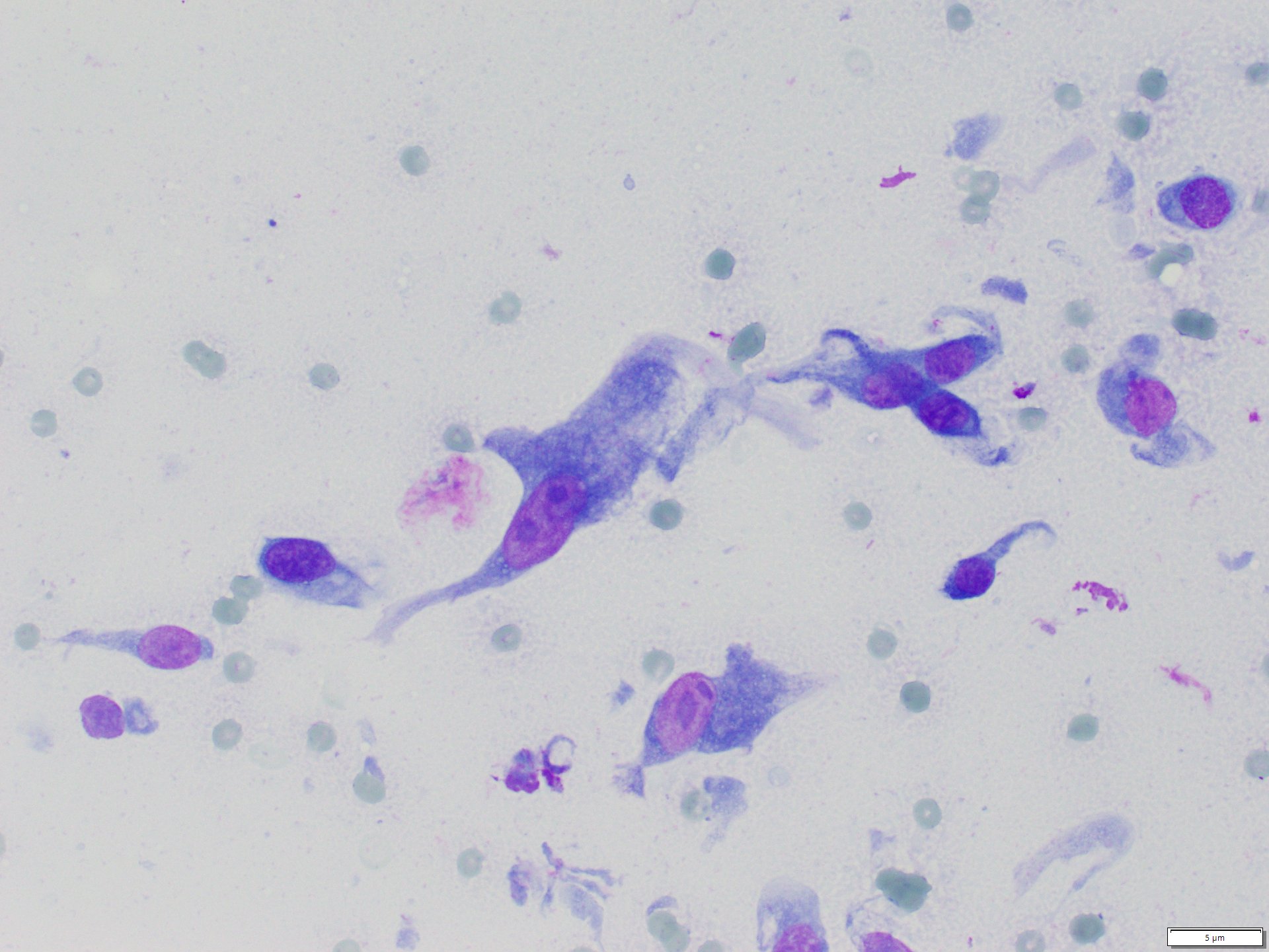
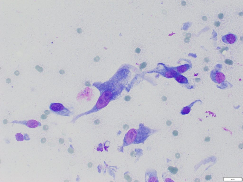
Of all the masses that occur in the skin and subcutis, probably the most care needs to be taken when evaluating spindle cells from mass lesions, as reactive spindle cells associated with fibroplasia cannot be easily distinguished from neoplastic cells on cytology. Often these lesions are firm and do not exfoliate well, making diagnosis even more problematic to make a definitive diagnosis on cytology.
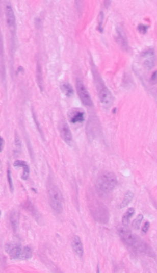
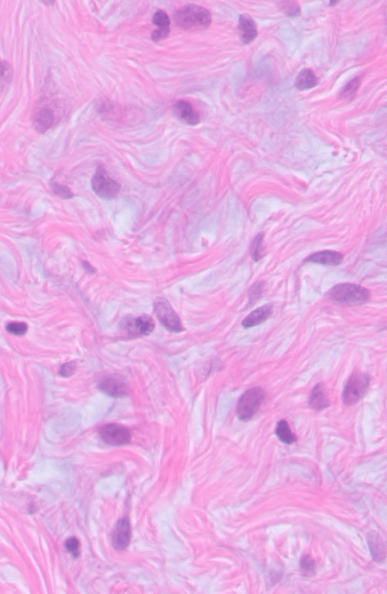
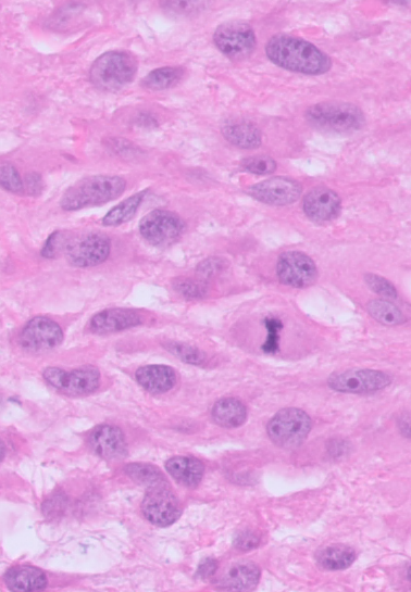
Moderate to large sized spindle cells can be present in areas of granulation tissue proliferation, fibrosis in lesions such as injection site and foreign body reactions, as well as spindle cell neoplasms. Confirming a lesion as a mesenchymal neoplasm therefore almost always requires histopathology. This is particularly true if any admixed inflammation is present. A history of progressive growth, infiltrative behaviour, and irregular mass borders, along with the lack of inflammatory cells, should increase suspicion of a neoplastic process.
Mesenchymal tumours can have benign (e.g. fibroma) and malignant (e.g. fibrosarcoma) forms and can have variable cell morphology in different areas of the mass. Unless marked cytologic atypia exists, making this distinction requires histopathology.
Reference:
Valenciano AC, Cowell RL. Cutaneous and Subcutaneous Lesions. In Cowell and Tyler’s (eds) Diagnostic Cytology and haematology of the Dog and Cat. 5th Edtn. Pp 74-100, Elsevier Inc., 2020.